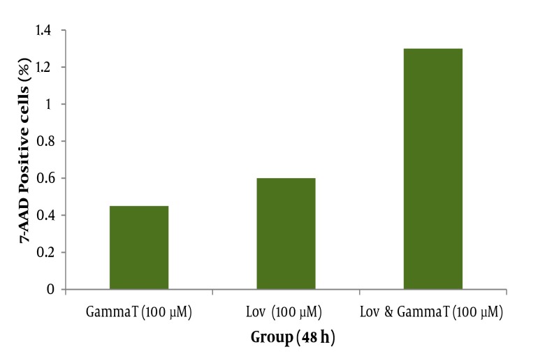
Figure 7. Late apoptosis positive cells determined by 7-AAD staining and flow cytometry in HT29 cell line. HT29 cells were grown in DMEM, exposed to specific agents and late apoptosis was determined by 7-AAD staining method. Far right group received 100μM γ-T and also lovastatin for 48h simultaneously.
* significant difference compared with γ-T (100μM,48h) and lovastatin (100μM, 48h) groups individually.

