Figure 1.
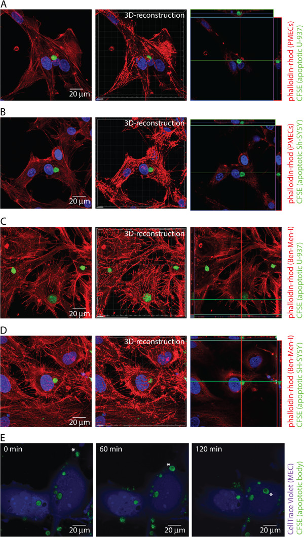
MECs take up apoptotic cells. Primary porcine MECs were incubated with CFSE-labeled apoptotic U-937 (A) or SH-SY5Y (B) cells for 4 h at a MEC:apoptotic body ratio of 1:5. Fixed MECs were permeabilized, actin was stained using phalloidine-rhodomine, and cells were analyzed by confocal microscopy. The right panel represents a 3D-reconstruction and a XYZ-view of the left panel. Ben-Men-I cells were incubated with CFSE-labeled apoptotic U-937 (C) or SH-SY5Y (D) cells for 4 h at a MEC:apoptotic body ratio of 1:5 and analyzed as above. (E) CellTrace Violet-labeled live Ben-Men-I cells were incubated with CFSE-labeled apoptotic U-937 cells and confocal images were taken at 0, 60, and 120 min. The pictures show a representative time course of apoptotic body uptake (marked with *) by MECs.

