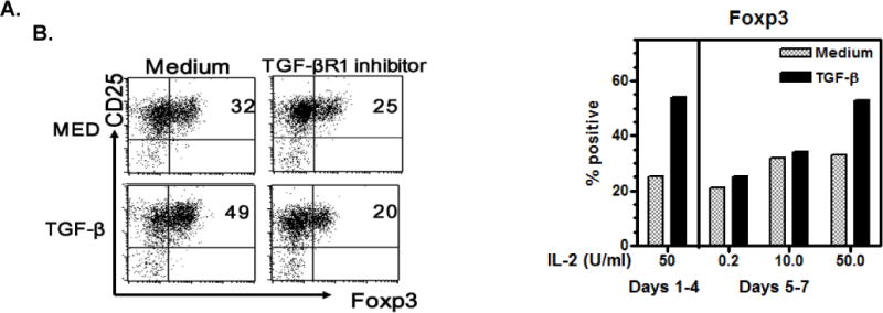
Figure 2. Foxp3 is expressed by activated human CD8+ cells, enhanced by TGF-β, and sustained by IL-2.

A. Naïve CD8 cells were stimulated with anti-CD3/28 coated beads (1 bead per 5 cells) with IL-2 (50U/ml) ± TGF-β1 (5ng/ml) and an alk5 TGF-βR1 signaling inhibitor (10μM) for 5 days. The cells were permeabilized, stained for Foxp3 and analyzed by flow cytometry. B. CD8 cells were stimulated as above with IL-2 50U/ml for 4 days, washed and IL-2 added back in the amounts indicated. Foxp3 was determined after culture for 2 more days. Here 50U/ml of IL-2 was required to sustain Foxp3 expression.
