
Figure 5. Characteristics of CD8regs induced with immobilized anti-CD3 and anti-CD28.
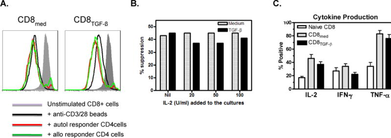
A. CD8regs are not anergic. The protocol described in Figure 4 was used for these experiments. After generation, CD8Medium and CD8TGF-β were labeled with CSFE and re-stimulated with anti-CD3/28 beads with or without autologous or allogeneic CD4responder cells. As shown, CD8Medium proliferate in response to secondary anti-CD3/28 stimulation, and this proliferation was enhanced when CD8TGF-β were when cultured with CD4 responder cells. The suppressive effects of these CD8regs on autologous and allogeneic CD4 responder cells are shown in Fig. 4B.
B. IL-2 does not inhibit suppressive activity. IL-2 was added in the concentrations shown to CD8regs mixed with CFSE-labeled responder CD4+ cells in suppressor assays in a ratio of 1:4. The experiment shown is representative of 4 similar experiments where IL-2 had no effect on CD8reg suppressive activity.
C. Comparison of cytokine production between CD8Medium and CD8TGF-β and unstimulated CD8+ cells. Using protocols described above, unstimulated CD8+ cells and those conditioned for 5 days were cultured with phorbol myristate acetate and ionomycin for 6 hours. Brefeldin A was added for the last 5 hours. The cells were permeabilized, stained for the cytokines shown, and intracellular cytokine production assessed by flow cytometry. In each of 6 experiments performed, the conditioned CD8+ cells produced more IL-2 and TNF-α than unstimulated CD8+ cells.