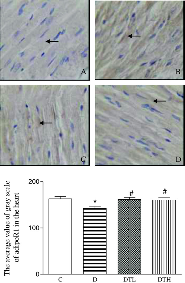
Figure 2.
Top panel: representative slides showing immunohistochemical staining of adiponectin receptor 1 (AdipoR1) (stained in brown as shown by arrow) in the myocardium. Slides A, B, C, D represent control, diabetic, diabetic treated with low dose (2 μg · kg−1.d−1) of exenatide and diabetic treated with high dose (10 μg · kg−1.d−1) of exenatide, respectively. Magnifications × 400. Bottom: bar graph shows quantitative analysis of myocardial adipoR1 expression in control, diabetic and diabetic treated with low (2 μg · kg−1.d−1) or high (10 μg · kg−1.d−1) dose of exenatide. The adiponectin receptor 1 (adipoR1) expression was quantified using the average value of gray scale which had an inverse proportion to the positive stained intensity. Control (C, n = 7), diabetic (D, n = 10), diabetic treated with low dose (2 μg · kg−1.d−1) of exenatide (DTL, n = 10), diabetic treated with high dose (10 μg · kg−1.d−1) of exenatide (DTH, n = 9). Data are expressed as mean ± S.E.M. *P < 0.05 different from control, #P < 0.05 different from diabetic.

