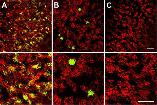
Figure 1.

Infection of well-differentiated BAEC by BPIV3 BRSV-GFP and BHV-1-GFP at different magnifications. A) BPIV3 was applied to the apical surface of ALI cultures (MOI~0.1) for 2 h. At 2 dpi, cultures were fixed and virus-infected cells were detected by immunostaining (green). B) BRSV-GFP was inoculated for 3 h and the cultures were fixed 3 dpi. Infected cells were detected by GFP expression. C) BHV-1-GFP was applied at an MOI of 0.1 to the apical side of ALI cultures for 2 h. 2 days later, cultures were fixed. Cilia were visualized by staining against β-tubulin (red). Scale bars = 50 μm.
