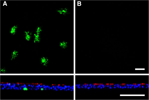
Figure 3.

BHV-1-GFP infection of the basolateral surface. A) Cells were infected at the basolateral surface by exposing the inverted filter membrane to BHV-1-GFP at an MOI of 1 for 2 h. B) As a control, inverted filters were infected by BRSV-GFP for 3 h. Infection was monitored 1 dpi (BHV-1-GFP) or 3 dpi (BRSV-GFP) by staining for β-tubulin (red) and DAPI (blue). Virus-infected cells were detected by GFP expression. Lower panels show vertical sections. Scale bars = 100 μm.
