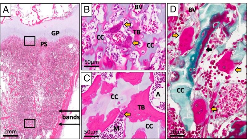
Figure 4.
Postdenosumab histopathology. A, An hematoxylin-eosin-stained section of the patient's growth plate taken 17 months after discontinuation of denosumab at the time of his second amputation showed sclerotic bands (bands with arrows) distant from the growth plate (GP). Calcified cartilage was present not only in the primary spongiosa (PS) but also within trabeculae in the region of the sclerotic bands. B, On high magnification of the osteochondral junction (upper box in panel A), calcified cartilage (CC) was found within the primary spongiosa. Also present were actively resorbing osteoclasts (yellow arrows) located adjacent to blood vessels (BV). C, Higher magnification of the bands (lower box in panel A) revealed islands of retained calcified cartilage (CC) within the trabecular bone (TB). An active osteoclast within a resorption pit can be seen in this area as well (yellow arrow). A, adipose tissue; M, normal marrow. D, A Goldner's trichrome-stained section taken from the same specimen at the osteochondral junction (upper box in panel A) again demonstrated the presence of calcified cartilage (CC) within the primary spongiosa. It also revealed actively resorbing osteoclasts (yellow arrows) and the presence of osteoid surfaces (black arrow), suggesting active bone formation and resorption.

