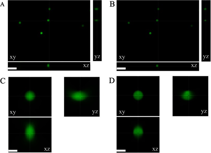
Figure 1.
Original and deconvolved wide-field microscope images of calibration microspheres. (A) An xy, xz, and yz view of 30% InSpeck Green fluorescent calibration microspheres imaged on a Zeiss Axiovert 200M microscope with a 100×/1.4-NA oil immersion lens and AxioCam high-resolution camera with mercury lamp power of 0.5% and exposure time of 120 ms. (B) Images as in A deconvolved with the blind deconvolution algorithm and the default settings in AutoQuant X3. (C and D) Zoomed-in images of one of the microspheres in A and B, respectively. Image display settings were adjusted independently so images appear to be of similar intensity, although deconvolved images have much higher intensities. Scale bars are 10 μm (A and B) and 3 μm (C and D).

