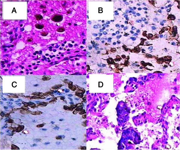
Figure 11.
Hematoxylin and eosin (H&E). A: The alveolar spaces were filled with many red blood cells and phagocytic cells with hemosiderin (heavy arrowhead). The alveolar walls were infiltrated by plasma cells (triangulate arrowhead) and lymphocytes (light arrowhead); 200×. Immunohistochemical staining, B: Gland epithelium in an alveolus, CK7+ (heavy arrowhead). Infiltrating plasma cells and lymphocytes in the alveolar wall, CK7- (light arrowhead), H&E 200×, C: Phagocytic cells in the alveolar space, CD68+ (heavy arrowhead). Plasma cells and lymphocytes in the alveolar walls, CD68- (light arrowhead), H&E 200×. D: Atypical tubular-gland structures of decidual lesions were detected in the alveolar space (light arrowhead). Structure of alveolar wall (heavy arrowhead); 100×.

