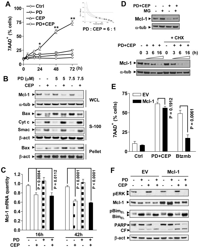
Figure 1. Combined treatment with CEP3891/PD184352 down-regulates Mcl-1 and induces apoptosis in MM cells including those ectopically over-expressing Mcl-1.
(A) U266 cells were co-exposed to 400 nM CEP3891±7.5 µM PD184352 for indicated interval, after which cell death was monitored by flow cytometry. Values represent the means and SD for three separate experiments performed in triplicate (* P<0.01 or ** P<0.001). U266 cells were treated (48 h) with a range of concentrations of CEP3891±PD184352 at a fixed ratio (6∶1), after which median dose effect analysis was used to characterize the nature of the interaction (inset). (B) U266 cells were treated with 400 nM CEP3891±PD184352 at the indicated concentrations for 16 h, after which subcellular fractions were prepared as discribed in meterial and method. Western blot analysis was performed using the indicated primary antibodies. WCL, whole cell lysate; S-100, cytosol; pellet, mitochondria-enriched fraction; Cyto c = cytochrome c. For these and all subsequent Western blot analyses, each lane was loaded with 20 µg of protein; blots were stripped and re-probed with α-tubulin (α-tub) or β-actin (β-act) antibodies to ensure equal loading and transfer; two additional studies yielded equivalent results. (C) U266 cells were treated with 400 nM CEP3891±7.5 µM PD184352 for 16 and 42 h, after which real-time qRT-PCR was performed to quantify Mcl-1 mRNA. Values represent the means and SD for three separate experiments. (D) U266 cells were incubated with 400 nM CEP3891±7.5 µM PD184352 in the presence or absence of 300 nM MG-132 (16 h, upper) or 1 µM CHX (3, 6, and 16 h, lower), after which protein levels of Mcl-1 were assessed by Western blot analysis. (E) U266 cells were stably transfected with a construct encoding human full-length Mcl-1 or empty vector (EV). Following treatment with 500 nM CEP3891±7.5 µM PD184352 for 48 h, the percentage of dead cells was determined by flow cytometry. In parallel, empty-vector and Mcl-1 over-expressing U266 were treated with 5 nM bortezomib as a control. Values represent the means and SD for three separate experiments performed in triplicate. (F) Alternatively, cells were subjected to Western blot analysis using the indicated primary antibodies. CF, cleavage fragment.

