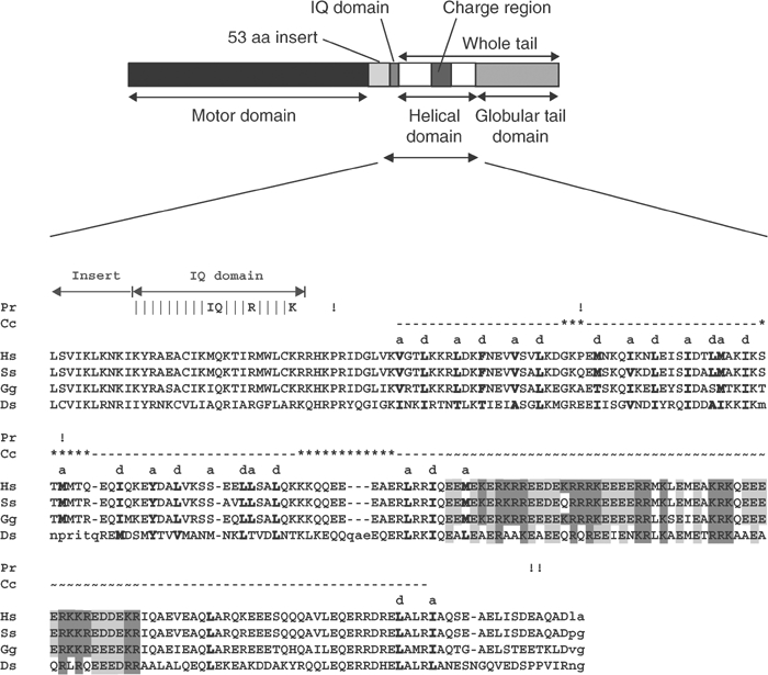
Figure 7.

Cartoon showing the different domains of myosin VI together with a sequence alignment and heptad repeat prediction for the myosin VI helical tail domains from different species. The cartoon shows the different domains of myosin VI and below is the sequence alignment of the helical tail domains (amino acids 812–1034) and their analysis using the COILS prediction program (Lupas, 1996). Vertical dashes show the IQ motif with the key residues labelled. The dashed line shows the presence of the putative coiled-coil sequence with hydrophobic a and d residues of the heptad repeat labelled and displayed in bold. Note: The complete sequence alignment includes several species not shown here; hence, hydrophobic residues that are not part of the heptad repeat but are highly conserved in the complete alignment are also shown in bold. Possible breaks in the putative coiled coil are shown by asterisks (***). An exclamation mark pinpoints helix breaking proline residues even if the proline is not conserved in all sequences. ∼∼∼ shows the highly charged region, and within this region, positively and negatively charged residues are shaded in dark and light grey, respectively. Accession numbers for myosin VI sequences are as follows: Hs (Homo sapiens), AAC51654; Ss (Sus scrofa), A54818; Gg (Gallus gallus), CAB96536; Ds (D. melanogaster), Q01989.
