Figure 5.
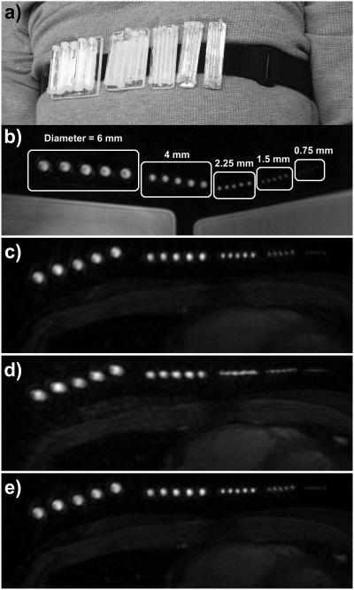
Free-breathing study with resolution phantom. (a) Photograph showing the resolution phantom strapped around the chest of a volunteer. The phantom consisted of five groups of five equally spaced vials, with inner diameters of 6, 4, 2.25, 1.5, and 0.75 mm. (b) An axial slice through the phantom is shown using a static 3D cones acquisition with the phantom strapped around a large doped-water phantom. The inner diameters of each vial are labeled. (c–e) A free-breathing 3D cones acquisition was carried out with the phantom strapped around the chest of a volunteer. An axial slice through the phantom is shown from images reconstructed with (c) no motion correction, (d) rigid-body translational motion correction using SI, AP, and RL trajectories derived from iNAVs located on the volunteer’s heart, and (e) autofocus motion correction using the same iNAV measurements as (d).

