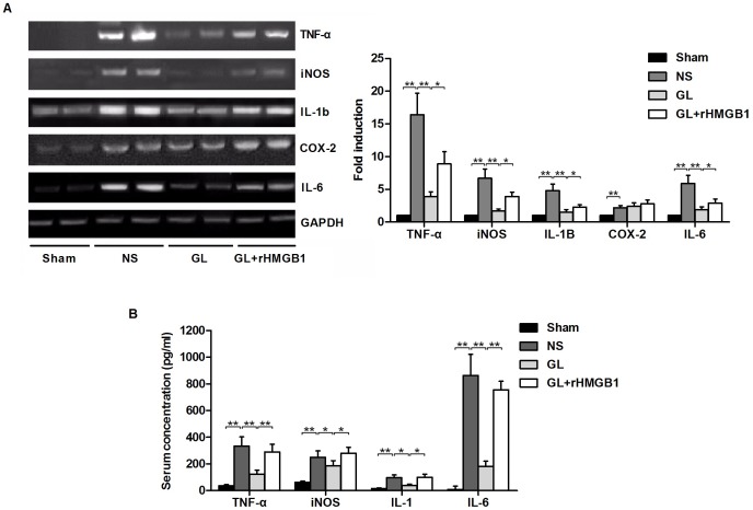
Figure 4. Expression of inflammation- and oxidative stress-related molecules in the brain and serum of MCA-occluded rats at 48 h after reperfusion.
A) Representative blots showing the effects of GL with or without rHMGB1 treatment on mRNA expression levels of pro-inflammatory and oxidative stress markers: TNF-α, iNOS, IL-1β, COX-2 and IL-6. GAPDH was used as a loading control. The bar graph showing semi-quantitative densitometric analysis summarises the fold change of TNF-α, iNOS, IL-1β, COX-2 and IL-6 expression in each group. B) Serum concentrations of TNF-α, iNOS, IL-1β, COX-2 and IL-6 at 48 h after I/R in each groups are determined. Values are means±SEM, n = 8 for each group. *P<0.05, **P<0.01 (t test).

