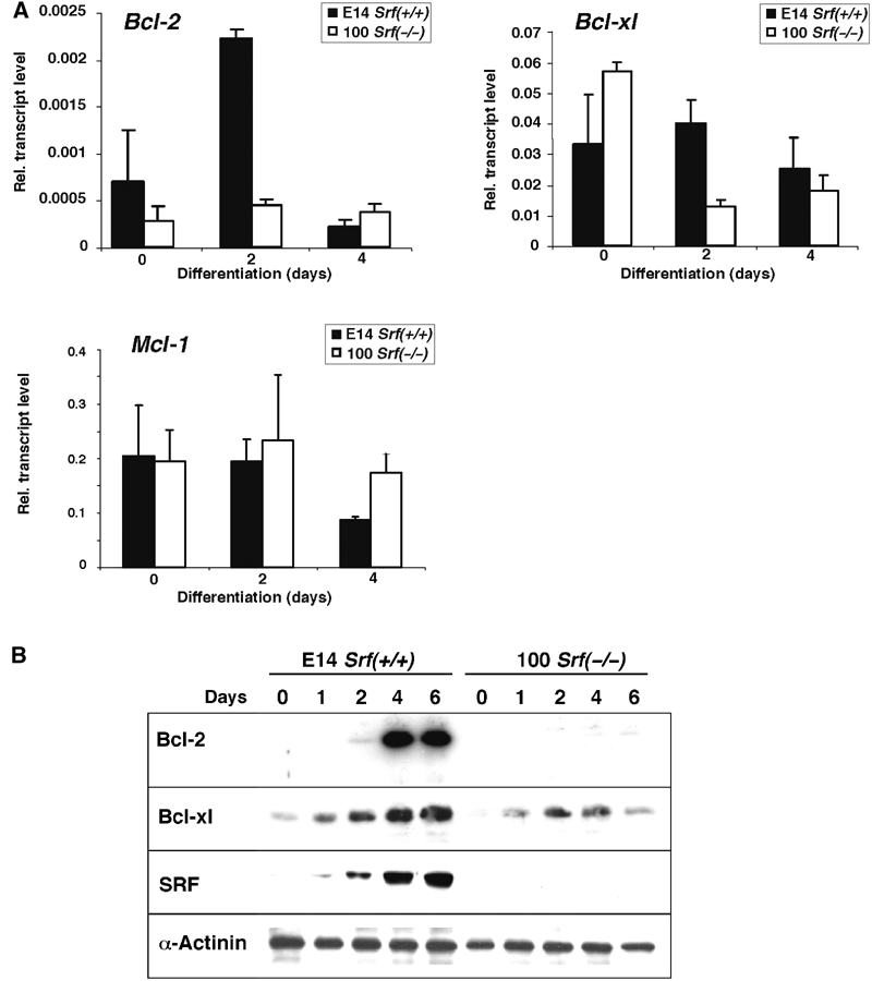
Figure 3.

Expression of antiapoptotic Bcl-2 family members in differentiating ES cells. (A) mRNA expression. Quantitative RT–PCR analysis was performed with RNA from E14 Srf(+/+) and 100 Srf(−/−) ES cells differentiated for 0, 2, and 4 days under monolayer conditions. Values represent the mean of three independent experiments ±s.d. (B) Expression of antiapoptotic Bcl-2 proteins. ES cell differentiation and extract preparation was carried out as described in Figure 1. Western blotting was performed using hamster anti-Bcl-2, mouse anti-Bcl-xl, and rabbit anti-SRF, as well as anti-α-actinin antibody for loading control.
