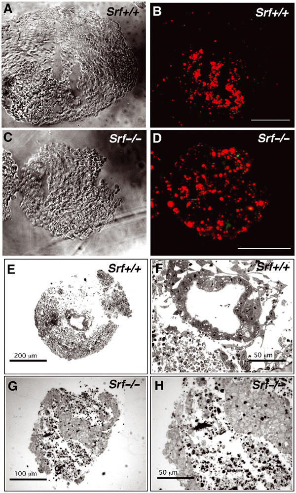
Figure 7.

EBs derived from Srf(−/−) ES cells display increased apoptosis and impaired cavity formation. (A–D) Increased, widespread apoptosis in day-5 Srf(−/−) EBs revealed by TUNEL staining (bar=100 μm). Note the size difference between Srf(+/+) and Srf(−/−) EBs. (E–H) Light microscopy on cryosections (10 μm) of day-8 EBs derived from either Srf(+/+) (E, F) or Srf(−/−) (G, H) ES cells. The results show representative examples of multiple EBs examined. Srf(+/+) EBs form cavities, which are lined by polarized columnar epithelial epiblast (CEE) cells (F). Srf(−/−) EBs display large areas of loosely arranged, fragmented cells, without forming distinct cavities (G). Note that a large fraction of cells inside Srf(−/−) EBs display condensed chromatin, a hallmark of apoptotic cell death (H).
