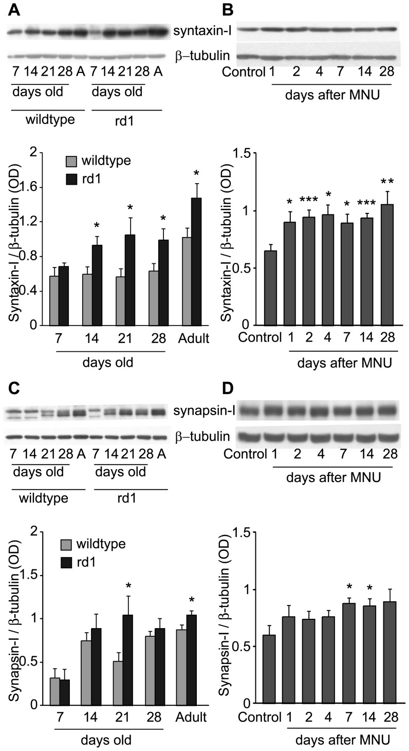
Figure 4. Amacrine cell-specific synaptic proteins were upregulated following photoreceptor loss.
A) Top: Representative blots of syntaxin-I and β-tubulin in retinas of wild-type and rd1 mice at different developmental stages (“A” is for “Adult”). Bottom: Ratio of syntaxin-I to β-tubulin for several animals (Mean ± SE). Syntaxin-I was upregulated in rd1 mouse retina from P-14 onwards (n = 7, 5, 7, 10, 14 for 7, 14, 21, 28 days old and Adult animals respectively). B) Top: Representative blots of syntaxin-I and β-tubulin in retinas of sham-injected control and at various days after MNU injection. Bottom: Ratio of syntaxin-I to β-tubulin for several animals (Mean ± SE). Similar to rd1 mouse, the levels of syntaxin-I were significantly higher in MNU-injected mice than in sham-injected controls. The upregulation was evident as early as one day after the injection (n = 10 for control; 9 for PID-1; 11 for PID-2, PID-4, PID-7 and PID-14; 10 for PID-28). C) Top: Representative blots of synapsin-I and β- tubulin in retinas of wild-type and rd1 mice at different developmental stages. Bottom: Ratio of synapsin-I to β-tubulin for several animals (Mean ± SE). Synapsin-I was upregulated in rd1 mouse from P-21 onwards till adult stage, although it was not statistically significant at P-28 (n = 4, 6, 8, 9 and 12 for 7, 14, 21, 28 days old and adult animals respectively). D) Top: Representative blots of synapsin-I and β-tubulin in retinas of sham-injected control and at various days after MNU injection. Bottom: Ratio of synapsin-I to β-tubulin for several animals (Mean ± SE). Similar to rd1 mouse, the levels of synapsin-I were significantly higher in MNU-injected mice than in sham-injected controls. The upregulation was evident from 7 days onwards after the injection (n = 7 for control; 6 for PID-1; 8 for PID-2, PID-4, PID-7 and PID-28; 9 for PID-14). *p<0.05; **p<0.01; ***p<0.005.

