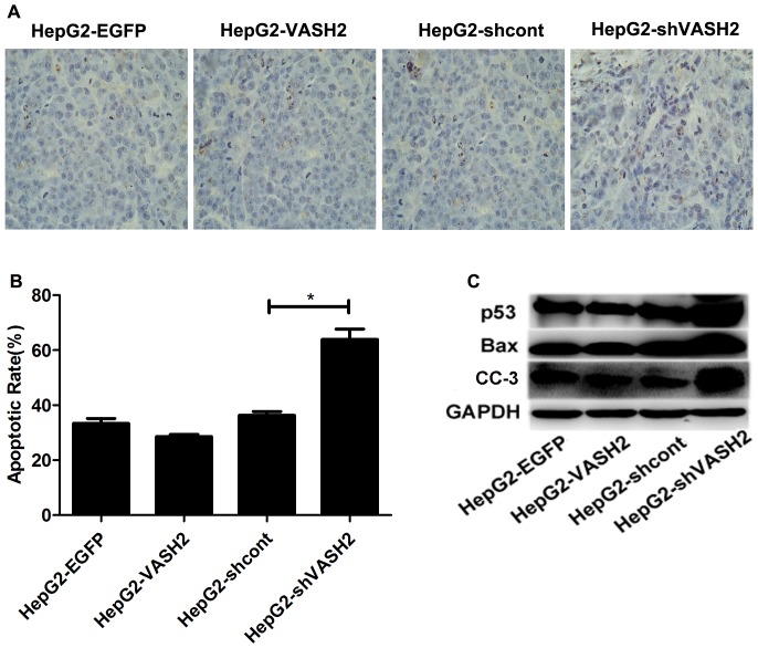
Figure 6. Knockdown of VASH2 enhanced the apoptosis of tumor cells by CDDP treatment.
(A and B) TUNEL analysis showing enhanced apoptosis rate in tumor cells when the HepG2-shVASH2 group was compared with the HepG2-shcont group (P<0.05). However, no statistical difference was found in HepG2-VASH2 vs. HepG2-EGFP (P>0.05) at 40× magnification. (C) Western blot analysis showed that p53, Bax, and CC-3 were highly expressed in the HepG2-shVSAH2 group.

