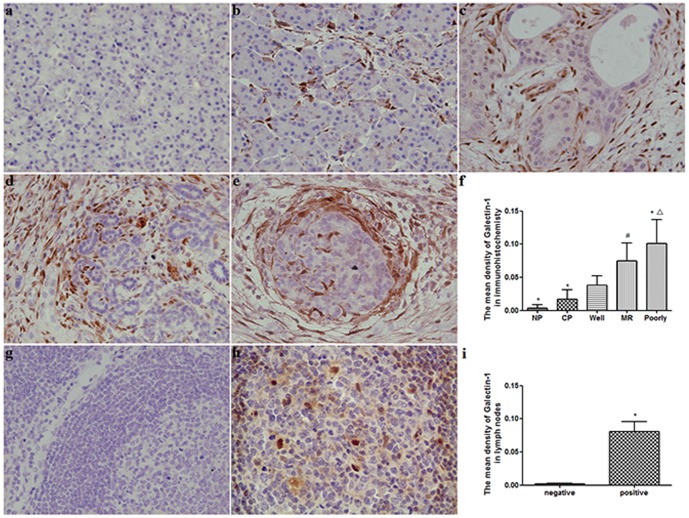
Figure 1. Characterization of Galectin-1 expressed in pancreatic disease.
(a) Negative Galectin-1 expression in normal pancreas tissue. (b) Weak Galectin-1 expression in the stromal of chronic pancreatitis tissue. (c–e) Strong Galectin-1 expression in stromal cells of pancreatic cancer tissue, and increased from well (c), moderately (d) to poorly (e) differentiated pancreatic cancer. (f) Quantification of mean density of Galectin-1 expression in different pancreatic tissues. *p<0.01 vs. well, # p<0.05 vs. well, △ p<0.05 vs. MR. NP, normal pancreas; CP, chronic pancreatitis; Well, Well differentiated pancreatic cancer; MR, moderately differentiated pancreatic cancer; Poorly, poorly differentiated pancreatic cancer. Original magnification: ×200. (g) Galectin-1 expression was negative in non-metastatic peripancreatic lymph nodes. (h) Galectin-1 positive staining was found in metastatic peripancreatic lymph nodes. (i) Quantification of mean density of Galectin-1 expression in non-metastatic and metastatic peripancreatic lymph nodes. *P<0.01 vs. negative.

