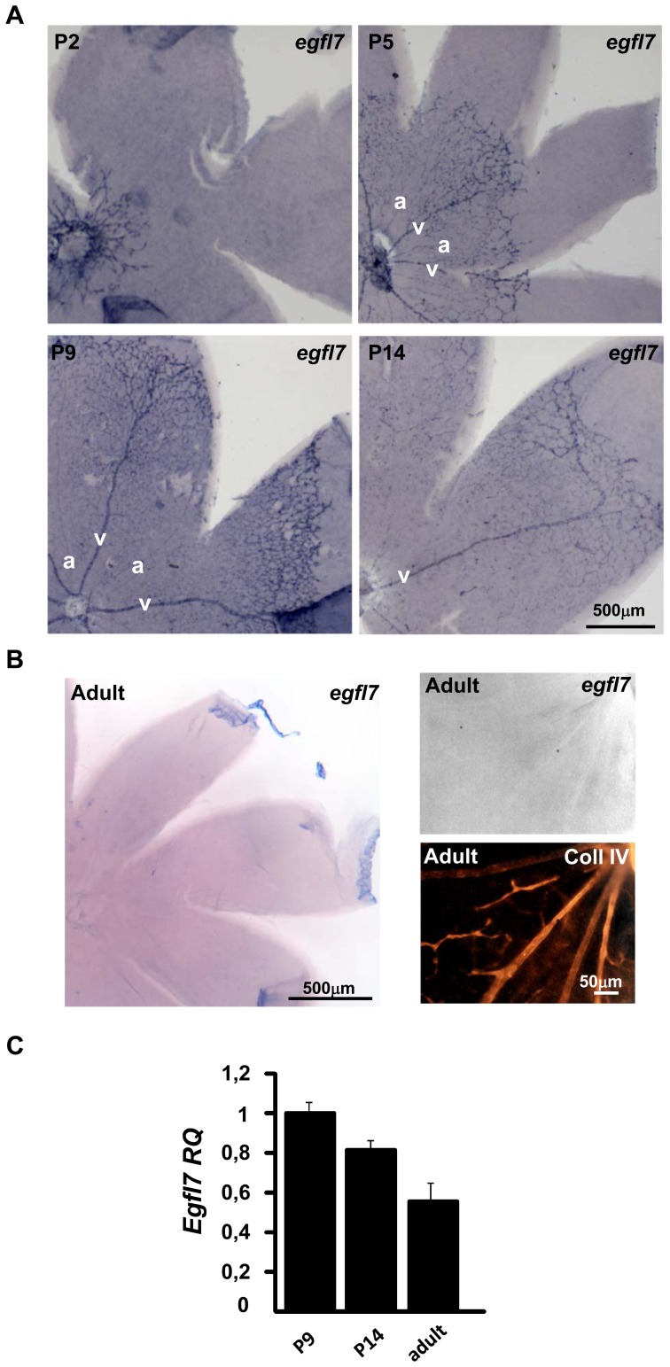
Figure 1. Egfl7 expression during retinal vascular development.
A: egfl7 expression detected by in situ hybridization (blue staining) in flat mounted retinas of two- (P2), five- (P5), nine (P9) and fourteen- (P14) day old mouse pups. B: Left panel: egfl7 expression detected by in situ hybridization in flat mounted adult retina. Right panel: higher magnification of the left panel; egfl7 expression detected by in situ hybridization (upper panel), collagen IV immunostaining of the same retina area (lower panel). C: Relative quantification by RT-qPCR of egfl7 transcripts in retina of nine- (P9), fourteen- (P14) day old mouse pups and adult mice. Levels were normalized to those of P9 retina arbitrarily set to 1. Magnifications are indicated.

