
Figure 1.
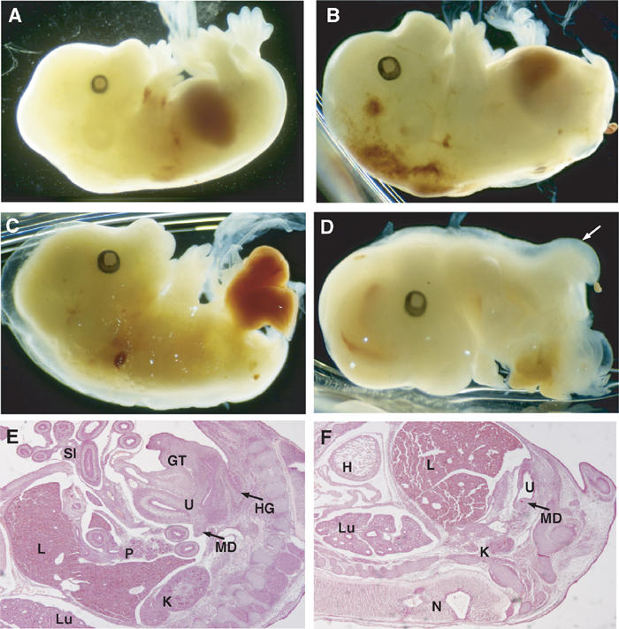
Tcf4−/−/Tcf1−/− embryos show severe caudal truncations. Lateral views of E14.5 embryos. (A) Tcf4+/−/Tcf1−/− and (B–D) Tcf4−/−/Tcf1−/− littermates. Panel B shows vestigial hindlimbs and the absence of a tail in mutant embryos. In more severe cases as depicted in panels C and D, internal organs such as the gut and liver are exposed. In several instances, the neural tube branched out in mutant embryos as shown in (D, F) (see arrow). Panels E and F represent haematoxylin and eosin stainings of sections of embryos depicted in (A, B), respectively. Various structures are identified as follows: (H) heart, (Lu) lungs, (L) liver, (K) kidneys, (U) urogenital sinus, (MD) mesonephric duct, (GT) genital tubercle, (SI) small intestine, (HG) hindgut and (N) neural tube.