
Figure 5.
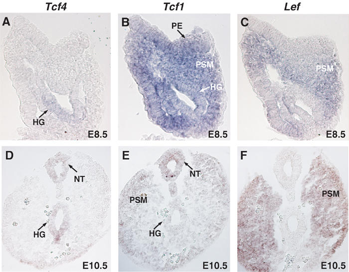
Comparison of Tcf4, Tcf1 and Lef expression. In situ hybridizations on sections of E8.5 and E10.5 wild-type embryos were performed using specific probes recognizing Tcf4 (A, D), Tcf1 (B, E) and Lef (C, F). Top panels A–C depict sections through the primitive streak region of E8.5 embryos, while bottom panels D–E show sections through the tail bud of E10.5 embryos. Various structures are identified as follows: (PE) primitive ectoderm, (NT) neural tube, (PSM) presomitic mesoderm and (HG) hindgut. Tcf4 was specifically expressed in the hindgut at E8.5 and E10.5 in the neural tube and hindgut. Tcf1 was found in all three germ layers at both E8.5 and E10.5. Lef was detected in the presomitic mesoderm in both stages examined and was absent from the primitive gut.