
Figure 6.
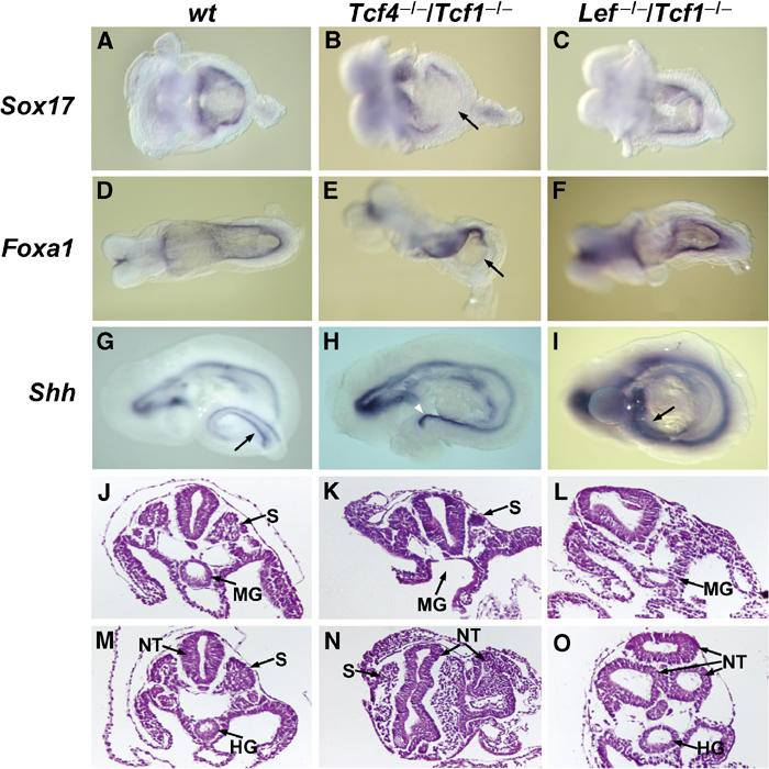
Impaired expansion of caudal endoderm in Tcf4−/−/Tcf1−/−embryos. Whole-mount in situ hybridizations of E8.5–9.5 littermates using endodermal markers and histology of E9.5 normal and mutant embryos. (A, D, G, J, M) Wild-type embryos, (B, E, H, K, N) Tcf4−/−/Tcf1−/−embryos and (C, F, I, L, O) Lef−/−/Tcf1−/− embryos. The arrows in (B) and (E) point to the loss of Sox17 and Foxa1 staining in the caudal endoderm of Tcf4−/−/Tcf1−/−, while staining is normal in Lef−/−/Tcf1−/− embryos. Arrows in (G) and (I) point to Shh expression in the hindgut of wild-type and Lef−/−/Tcf1−/− embryos. The notochord in Tcf4−/−/Tcf1−/−embryos appears exposed due to the absence of an underlying gut tube (arrowhead in (H)). Panels J–L and M–O represent haematoxylin and eosin stainings of sections through the midgut region and hindgut, respectively. Various structures are identified as follows: (S) somites, (NT) neural tube, (MG) midgut and (HG) hindgut. Note that in panel K Tcf4−/−/Tcf1−/− embryos maintain somites but the primitive gut tube has not closed. Similar sections in Lef−/−/Tcf1−/− embryos show a properly formed gut tube and the absence of somites (panel L). The caudal sections (M–O) also reveal the presence of ectopic neural tissue in both Tcf4−/−/Tcf1−/− and Lef−/−/Tcf1−/− embryos.