
Figure 7.
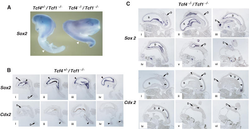
Anteriorization of the gastro-intestinal tract in Tcf4−/−/Tcf1−/− embryos. (A) Whole-mount in situ hybridizations of dissected anterior gastro-intestinal tracts of E14.5 Tcf4+/−/Tcf1−/− (left) and Tcf4−/−/Tcf1−/− (right) embryos using a stomach marker, Sox2. The gastro-intestinal tract of the normal embryo was severed to remove the intestinal tube. The truncated gastro-intestinal tract of Tcf4−/−/Tcf1−/− embryos is shown in its entirety. As shown by the arrowheads, Sox2 expression is confined to the stomach in normal littermates, while ectopic expression in the duodenum is apparent in mutants. (B) In situ hybridizations on consecutive sections (i–iv) of a single E13.5 Tcf4+/−/Tcf1−/− gastro-duodenal preparation. The top and bottom rows of panels were stained for Sox2 (stomach) and Cdx2 (intestine), respectively. The top and bottom sections i–iv represent equivalent regions and should be compared with each other. The arrowheads indicate regions of positive staining for both probes. (C) In situ hybridizations on consecutive sections (i–vi) of a single E13.5 Tcf4−/−/Tcf1−/− gastro-duodenal preparation. The top and bottom sets of panels were stained for Sox2 and Cdx2, respectively. Sections i–vi represent equivalent regions and should be compared with each other. Various structures are identified as follows: (S) stomach, (D) duodenum. The arrowheads in panels stained for Cdx2 represent regions of the duodenum that are devoid of Cdx2 expression but show high expression of Sox2. Note how the duodenum in mutants appears dilated compared to normal littermates (B). In addition, Cdx2 transcripts are confined to a small portion of the duodenum, and the remaining tissue expresses Sox2. Altogether, these data provide evidence for the occurrence of an anterior transformation in the gastro-intestinal tract of Tcf4−/−/Tcf1−/− embryos.