
Figure 4.
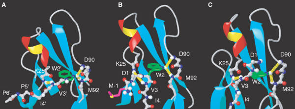
Conformation of the N-terminal region of ECAD12 in different crystal forms. (A) Details of the crystal structure of dimeric ECAD12 (1q1p) showing the docking of W2 into the pocket of the partner molecule. Residues that stem from the symmetry equivalent molecule are marked by an apostrophe ('). The same region is shown for the crystal structures of M-ECAD12 (B, 1ff5) (Pertz et al, 1999) and human ECAD1 in complex with internalin (C, 1o6s) (Schubert et al, 2002). Long-range NOEs observed between W2Hɛ↔D90HN/M92HN and V3HN↔K25HN for the monomeric form of ECAD12 (see text and Figure 3C and D) are highlighted by dashed yellow cylinders in all crystal forms (A–C).