
Figure 5.
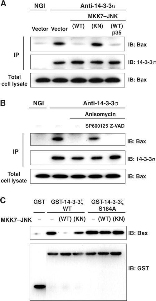
JNK promotes dissociation of Bax from 14-3-3 proteins. (A) HCT116 cells were transfected for 24 h with expression vectors for GFP, MKK7–JNK(WT or KN) and p35, as indicated. Cell lysates were then subjected either to immunoblot analysis with antibodies to Bax or to immunoprecipitation (IP) with antibodies to 14-3-3σ (or to normal goat IgG (NGI) precipitation); the resulting precipitates were subjected to immunoblot analysis with antibodies to 14-3-3σ and to Bax. (B) HCT116 cells were incubated first for 30 min with or without 20 μM SP600125 or 100 μM Z-VAD-CH2DCB and then for 3 h in the presence or absence of anisomycin (10 μg/ml). Cell lysates were then subjected to immunoprecipitation and immunoblot analysis as described in (A). (C) Equal amounts of recombinant GST–14-3-3ζ (WT or Ser184 → Ala mutant) or GST alone were incubated with or without recombinant MKK7–JNK(WT or KN) in the presence of ATP for 30 min, and the GST proteins were precipitated with glutathione-sepharose beads. The beads were then incubated with HeLa cell lysates, and subjected to immunoblot analysis with antibodies to Bax or to GST.