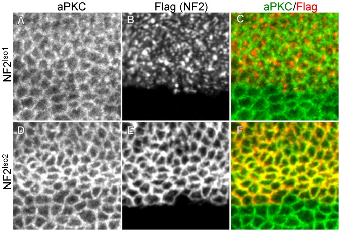
Figure 3. NF2 isoforms 1 and 2 display distinct localizations within the apical domain.
Wing imaginal discs expressing Flag-tagged NF2Iso1 (A–C) or NF2Iso2 (D–F) under the ApGal4 driver. Images are taken along the dorsal-ventral boundary to include the expression boundary of ApGal4, thereby demonstrating the specificity of anti-Flag staining in these tissues. Comparison to aPKC, a marker for the marginal zone in these cells (A,D), demonstrates that NF2Iso1 (B–C) is expressed in the apical cell cortex but is not closely associated with the marginal zone. In contrast, NF2Iso2 (E–F) is highly concentrated in the marginal zone together with aPKC.

