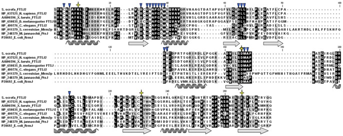
Figure 2. Protein sequence alignment of E. coli RrmJ with its FTSJ2 orthologs in 7 different species.
The α-helices and β-strands were based on the RrmJ protein structure (PDB code: 1EIZ). The stars and triangles indicate the K-D-K-E catalytic center and the SAM binding residues in RrmJ, respectively. The residues with identical and similar chemical properties are highlighted in black and gray, respectively.

