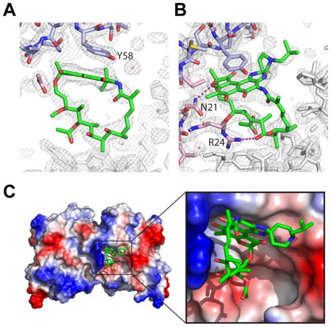
Figure 3. Crystal structure of rifabutin and BCL6-POZ domain.

Electron density corresponding to rifabutin, following refinement, in the context of surrounding electron density demonstrating the proximity of (A) tyrosine58 from one monomer of the POZ dimer and (B) asparagine21 and arginine24 from the other monomer. (C) Surface representation of BCL6-POZ with basic residues (including asparagine21 and arginine24) in blue and acidic residues in red. The napthoquinone rings of rifabutin are in proximity to tyrosine58 whilst the aliphatic bridge is adjacent to the basic surface.
