Figure 2.
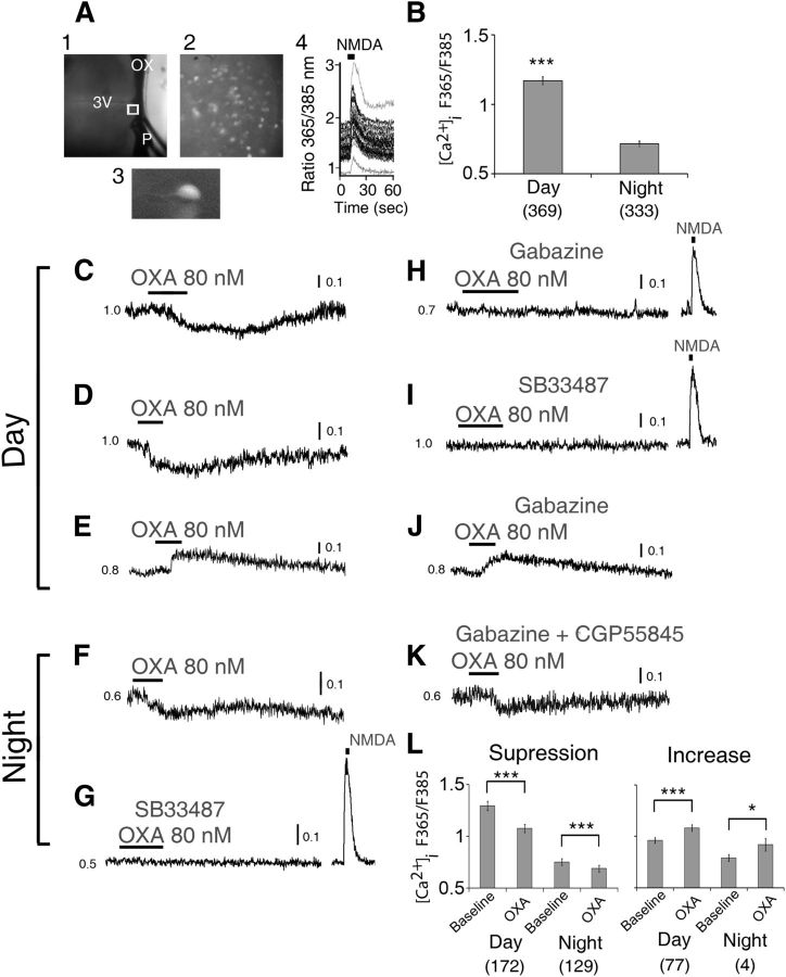
OXA mainly suppresses intracellular Ca2+ levels in SCN neurons. A, Simultaneous [Ca2+]i measurements in multiple SCN neurons in a hypothalamic slice, with (1) recording setup with pressurized puffer pipette (P) used for brief (5 s) focal application of NMDA; (2) SCN neurons (40×) loaded with LeakRes fura-2 AM (taken from region in (1) marked with a white box); (3) enlarged single LeakRes fura-2 AM loaded SCN neuron; (4) representative SCN neurons showing transient [Ca2+]i increase in response to a focal application of NMDA (200 μm, 5 s; duration black bar at top) in the late day (ZT10). B, Overall summary of measured baseline [Ca2+]i level in SCN cells showing significantly higher [Ca2+]i value during the day (ZT4–10) than at night (ZT16–22). During the day, OXA (80 nm) significantly suppressed (C, D; n = 172/249) or elevated (E; n = 77/249) the baseline [Ca2+]i ratio in 69 and 31% of responsive SCN neurons, respectively. At night, OXA decreased (F) the baseline [Ca2+]i ratio in 97% (n = 129/133) of responsive SCN neurons while increasing this ratio in only 3% (n = 4/133) of cells (data not shown). This nighttime effect of OXA on cytosolic calcium levels in SCN neurons was insensitive to coapplication of gabazine and CGP5585 (K), GABAA, and GABAB receptor antagonists, respectively, but was abolished when OXA receptor antagonist SB33487 was coapplied with OXA (G). In contrast, in the day, gabazine abolished OXA's suppression of cytosolic calcium levels in SCN cells (H), but not its induced elevation (J) of the ratio. G–I, In the presence of gabazine and SB33487 the cells showed an acute increase in their cytosolic calcium levels in response to a 200 μm focal application (5 s) of NMDA. L, Summary of OXA effects on [Ca2+]i level in SCN cells over the day and at night. All data shown are expressed as mean ± SEM; *p < 0.05,***p < 0.0001. Number of cells measured is shown in brackets. OX, optic chiasm; 3V, third ventricle; P, tip of the pressurized puffer pipette manifold.

