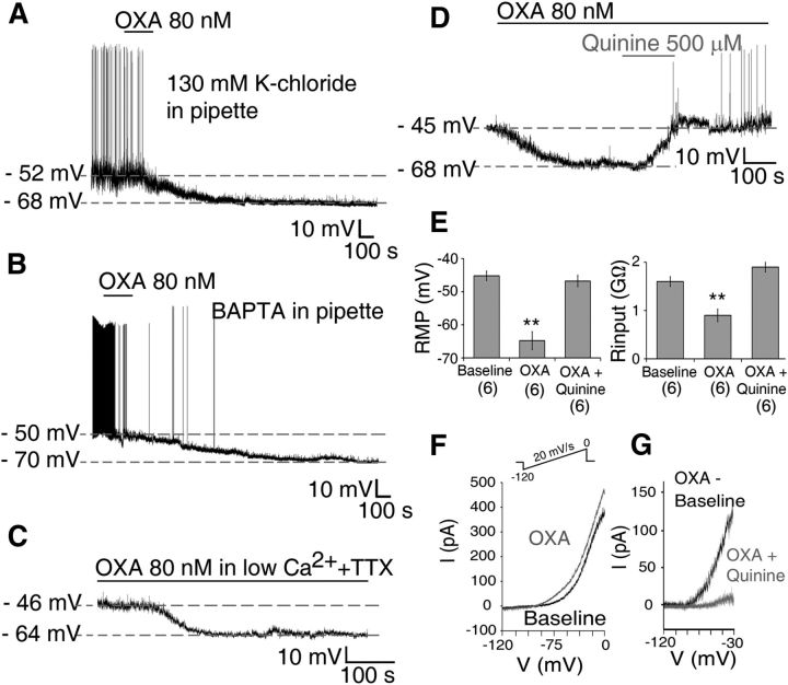
Figure 6.
OXA causes direct membrane hyperpolarization and suppression of Per1-EGFP neuronal activity at night. A–C, Bath application of OXA hyperpolarized Per1-EGFP neurons, and in all cells this hyperpolarization was significantly larger in duration (>50 min) at this circadian phase than during the day. A, B, OXA-induced suppression of Per1-EGFP cells persisted when (A) intracellular K+-gluconate was substituted with equimolar KCl solution; (B) intracellular Ca2+ solution was clamped at <90 nm with BAPTA-Ca2+ mixture, preventing Ca2+ mobilization from intracellular stores; and (C) when Ca2+ influx was prevented with low Ca2+/high Mg2+ extracellular solution. D, Bath application of quinine prevented OXA-hyperpolarizing effect. E, Summary of results obtained from six current-clamp cells where OXA-induced membrane hyperpolarization and accompanied reduction in Rinput were blocked when OXA was coapplied with quinine. F, I–V relationship during baseline (black trace) and in the presence of OXA (gray trace) obtained by slowly ramping the membrane voltage (see Materials and Methods and inset). G, Net OXA current obtained by subtracting OXA I–V from control I–V (black trace), with quinine potently inhibiting this OXA-induced current (gray trace). **p < 0.01. Data are expressed as mean ± SEM. Number of cells measured is shown in parentheses.

