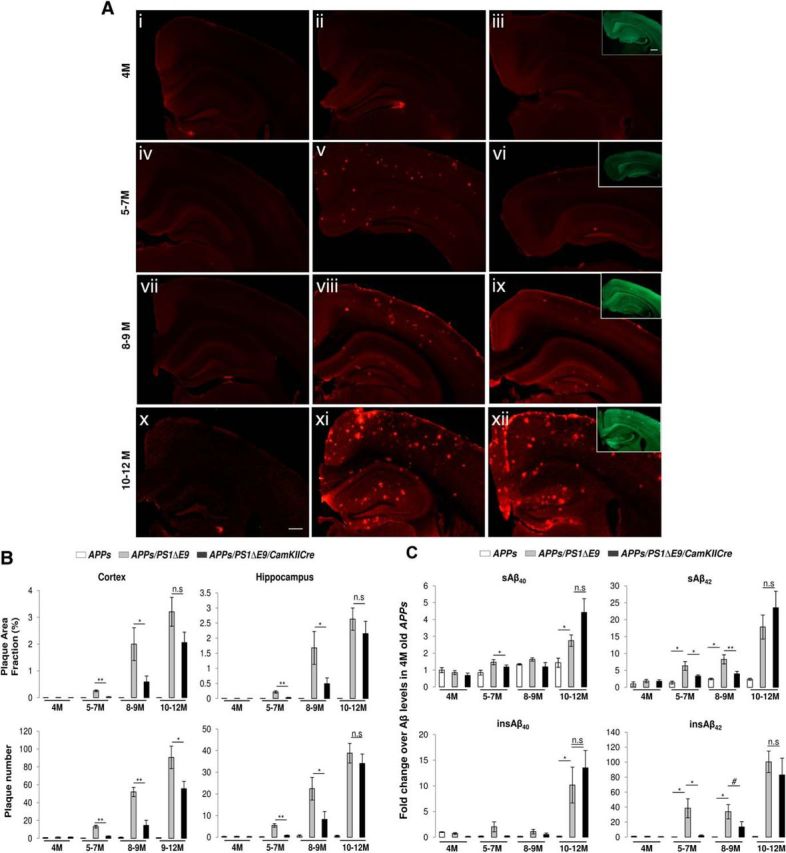
Figure 2.

Amyloid deposition and Aβ levels as a function of age in brains of APPswe, APPswe/PS1ΔE9flox, or APPswe/PS1ΔE9flox/CaMKIICre mice. A, Extent of amyloid deposition after postnatal ablation of FAD-linked mutant PS1 expression in mature excitatory neurons. Images of hemibrain sections from APPswe (Ai, Aiv, Avii, Ax), APPswe/PS1ΔE9flox (Aii, Av, Aviii, Axi), or APPswe/PS1ΔE9flox/CaMKIICre (Aiii, Avi, Aix, Axii) mice aged to 4, 5–7, 8–9, or 10–12 months immunostained with 3D6 (red). cGFP expression as observed by cGFP fluorescence (green) in APPswe/PS1ΔE9flox/CaMKIICre mice are shown within the insets in Aiii, Avi, Aix, and Axii. Scale bar, 500 μm. B, Quantification of amyloid deposition after postnatal ablation of FAD-linked mutant PS1 expression in mature excitatory neurons. Histograms show the amyloid plaque area fraction (percentage) and plaque numbers in the cortex or hippocampus of APPswe, APPswe/PS1ΔE9flox, or APPswe/PS1ΔE9flox/CaMKIICre mice aged to 4, 5–7, 8–9, or 10–12 months. n = 6 (4 months, APPswe), 7 (4 months, APPswe/PS1ΔE9flox), 6 (4 months, APPswe/PS1ΔE9flox/CaMKIICre), 5 (5–7 months, APPswe), 8 (5–7 months, APPswe/PS1ΔE9flox; plaque area fraction in the cortex and hippocampus was 0.255 ± 0.033 and 0.218 ± 0.039%, respectively, and the plaque number was 13.15 ± 1.49 and 5.47 ± 0.79, respectively), 8 (5–7 months, APPswe/PS1ΔE9flox/CaMKIICre), 3 (8–9 months, APPswe), 6 (8–9 months, APPswe/PS1ΔE9flox; plaque area fraction was 1.99 ± 0.61 and 1.67 ± 0.54%, respectively, and plaque numbers were 51.96 ± 5.18 and 22.4 ± 5.24, respectively), 7 (8–9 months, APPswe/PS1ΔE9flox/CaMKIICre; plaque area fraction in the cortex and hippocampus was 0.589 ± 0.21 and 0.49 ± 0.19%, respectively, and plaque numbers were 14.57 ± 5.64 and 8.22 ± 3.64, respectively), 3 (10–12 months, APPswe), 4 (10–12 months, APPswe/PS1ΔE9flox; plaque area fraction and plaque numbers in cortex were 3.2 ± 0.53% and 90.7 ± 12.6, respectively, and the plaque area fraction and plaque numbers in hippocampus were 2.62 ± 0.36% and 38.85 ± 4.55, respectively), and 5 (10–12 months, APPswe/PS1ΔE9flox/CaMKIICre; plaque area fraction and plaque number in the cortex were 2.05 ± 0.39% and 55.6 ± 8.36, respectively, and the plaque area fraction and plaque numbers in the hippocampus were 2.15 ± 0.41% and 34.08 ± 4.35, respectively). Plaque area fraction in the cortex: *p = 0.021 for8–9 months and **p = 0.00013 for 5–7 months for APPswe/PS1ΔE9flox versus APPswe/PS1 ΔE9flox/CaMKIICre groups. Plaque numbers in the cortex: *p = 0.023 for 10–12 months,**p < 0.0001 for 5–7 months, and **p = 0.008 for 8–9 months for APPswe/PS1ΔE9flox versus APPswe/PS1ΔE9flox/CaMKIICre groups. Plaque area fraction in the hippocampus: *p = 0.025 for 8–9 months and **p = 0.001 for 5–7 months for APPswe/PS1ΔE9flox versus APPswe/PS1ΔE9flox/CaMKIICre groups. Plaque numbers in the hippocampus: *p = 0.022 for 8–9 months and **p = 0.0004 for 5–7 months for APPswe/PS1ΔE9flox versus APPswe/PS1ΔE9flox/CaMKIICre groups. n.s., Not significant. C, Changes in steady-state levels of Aβ40 and Aβ42 peptides after postnatal ablation of FAD-linked mutant PS1 expression in mature excitatory neurons. Histograms show fold changes in sAβ40, sAβ42, insAβ40, or insAβ42 levels in hemibrains of APPswe, APPswe/PS1ΔE9flox, or APPswe/PS1ΔE9flox/CaMKIICre mice aged to 4, 5–7, 8–9, or 10–12 months over the levels observed in age-matched APPswe mice group. n = 6 (4 months, APPswe), 7 (4 months, APPswe/PS1ΔE9flox), 7 (4 months, APPswe/PS1ΔE9flox/CaMKIICre), 5 (5–7 months, APPswe), 8 (5–7 months, APPswe/PS1ΔE9flox), 8 (5–7 months, APPswe/PS1ΔE9flox/CaMKIICre), 2 (8–9 months, APPswe), 5 (8–9 months, APPswe/PS1ΔE9flox), 6 (8–9 months, APPswe/PS1ΔE9flox/CaMKIICre), 3 (10–12 months, APPswe), 5 (10–12 months, APPswe/PS1ΔE9flox), and 5 (10–12 months, APPswe/PS1ΔE9flox/CaMKIICre). * p < 0.05, **p < 0.01, #p = 0.058. n.s., Not significant.
