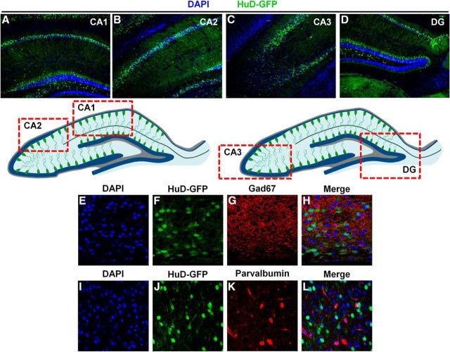
Figure 2.
HuD is not expressed in Gad67 or parvalbumin+ interneurons and is expressed in the CA1–3 and dentate gyrus of the hippocampus. A–D, Representative 10× confocal images of hippocampal subregions CA1, CA2, CA3, and dentate gyrus, respectively. Bottom, Schematic of HuD-GFP expression in the hippocampus. Red boxes denote regions where representative confocal images were captured. E–H, Representative 60× confocal images of cortical of DAPI (blue), HuD-GFP (green), Gad67 (red), and merged channels, respectively. I–L, Representative 60× confocal images of cortical of DAPI (blue), HuD-GFP (green), parvalbumin (red), and merged channels, respectively.

