Figure 7.
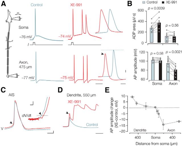
Kv7 channels differentially regulate back-propagating and forward-propagating action potentials. A, Left, Schematic of the simultaneous soma–axon whole-cell recording. Right, Action potentials evoked by brief (3 ms) somatic current injection simultaneously recorded at an axonal recording distance of 475 μm in control condition (gray) and after bath application of XE-991 (10 μm, red). Note the additional high-frequency spikes in the presence of XE-991. Calibration: 10 ms. High magnification of the same traces shows the reduced axonal action potential amplitude (arrow). Calibration: 0.5 ms, 10 mV. B, Bar graphs and individual experiments of the area under the curve reveal an increase in the somatic ADP (n = 9) after XE-991 application but decrease in the distal axons (>150 μm, n = 9). C, Action potentials were evoked at the soma and simultaneously recorded from the AIS. The AIS action potential threshold was significantly increased (n = 5, p = 0.022, arrow). Inset, The phase plot (time derivative vs voltage) highlighting the voltage threshold. Calibration: 0.5 ms, 10 mV (inset: 5 mV, 0.1 kV s−1). D, XE-991 block in the dendrite increased the action potential amplitude (arrow). In this example, the dendrite generated a long-lasting plateau depolarization associated with high-frequency burst firing at the soma. Calibration: 10 ms, same voltage scaling as in C. E, Summary data of all dual whole-cell recordings (n = 25) showing the location dependence of action potential change in XE-991. Action potential amplitudes increase in the distal dendrite, are maintained at the soma, but are substantially reduced in the axon. Symbols represent x and y mean ± SEM.

