Figure 2.

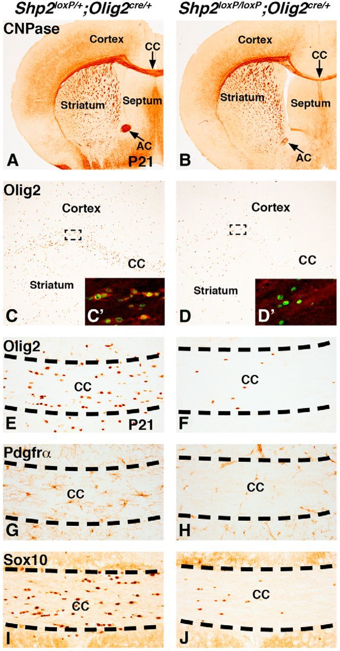
Reduced OLs and OPCs in Shp2 cKOs at P21. CNPase expression in OLs is reduced in the postnatal white matter regions (CC and AC) in Shp2 cKOs (Shp2loxP/loxP;Olig2cre/+) (B) compared with controls (Shp2loxP/+;Olig2cre/+) (A). Olig2 expression labels both OLs and OPCs at P21 (C) and is generally reduced in Shp2 cKOs (D). Dashed lines indicate representative areas for C and D insets that show Shp2 (red) and Olig2 (green) double stains. Shp2 staining in the cytoplasm is detected in Olig2-positive cells (C′) in controls. However, the few remaining Olig2 cells in Shp2 cKOs do not show any Shp2 staining in the cytoplasm (D′). Olig2-, Pdgfrα-, and Sox10-positive cells are severely reduced in the medial CC of Shp2 cKOs (F, H, and J, respectively) compared with controls (E, G, and I, respectively).
