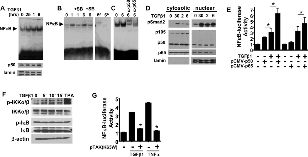
Figure 2. TGFβ1 induces nuclear NFκB binding activity and enhances p50 and p65 driven gene expression.
(A) Top: NFκB EMSA of nuclear extracts from primary keratinocytes treated with TGFβ1(1 ng/mL) for the indicated times. Bottom: immunoblot of nuclear extracts showing equal amounts of NFκB p50 and lamin loading control (B) NFκB EMSA of nuclear extracts from primary keratinocytes treated with TGFβ1 for the indicated times +/− the ALK5 inhibitor SB431542 (1 uM). Extracts isolated 6h post TGFβ1 treatment were incubated with labeled mutant NFκB oligo (6a) or unlabelled wildtype NFκB oligo(6b). (C) NFκB EMSA of TGFβ1 treated 6 hr nuclear extracts incubated with anti-p50 and anti-p65 antibodies. (D) Immunoblot of NFκB subunits and phospho-Smad2 levels in nuclear and cytosolic lysates from primary keratinocytes treated with TGFβ1. 50 g of nuclear extract was used to detect p65 in the nucleus. Cytoplasmic and nuclear lysates were run on the same gel, with intervening lanes removed. (E) Effect of pCMV-p50 or pCMV-p65 cotransfection on TGFβ1 induced NFκB-luc activity. All experiments were averaged from N=6 wells repeated 3 times. * indicates significant difference between indicated groups, p<0.05 (F) Immunoblots of phospho and total IKKα/β, phospho and total IκB in cytosolic extracts of TGFβ1 treated primary keratinocytes. Repeated 3 times. (G) pNFκB-luc reporter activity in primary mouse keratinocytes treated with or without TGFβ1 (1 ng/mL), TNFα (10 ng/mL) in the presence of TAK1K63W or control vector. Average of 3 wells, repeated twice. * indicates significantly different from TGFβ1 alone, p<.05.

