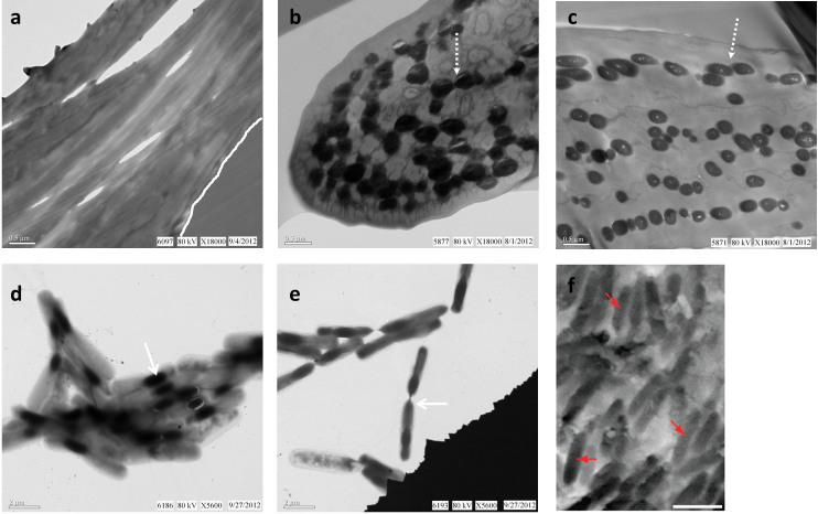
Figure 1. Electron micrographs of modern Gallus gallus feathers and Bacillus cereus pure culture, compared with an SEM image of a fossil feather from published work.
Chicken feathers were sectioned, stained, and viewed in transmission EM as described (see Methods). Melanosomes are observed (dashed arrow) in brown (b) and black (c) feathers but are absent in similarly prepared white feathers (a) (contact between feather and embedding medium delineated by white line in (a)). (d) Aggregation of B. cereus cells containing electron opaque internal endospores (arrow). (e) Two endospore-containing B. cereus cells aligned and connected (arrow), prepared and stained as described (see Methods). (f) SEM image of isolated feather of Jurassic bird Archaeopteryx with “[…] melanosomes (arrows) preserved […] as moulded imprints” (scale bar: 1 μm). Reprinted by permission from Macmillan Publishers Ltd: [NATURE COMMUNICATIONS]7, copyright (2012).

