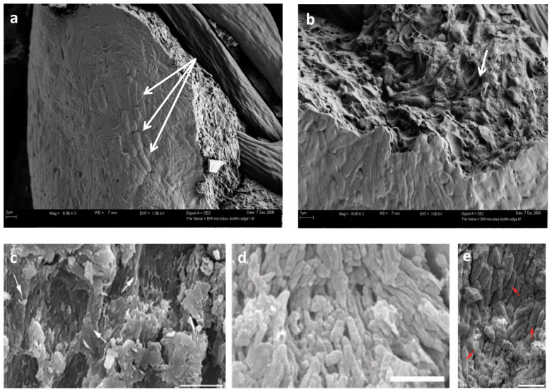
Figure 2. FE-SEM micrographs of biofilm overgrowth of extant feathers compared with published images of fossil feathers.
(a) Guineafowl feathers exposed to naturally occurring culture show strong alignment of microbial cells (arrows). (b) Higher magnification of biofilm edge in (a) showing where bacteria cells have been eliminated from the surrounding matrix (arrow), leaving voids similar to those figured in (c), which were identified as “[…] eumelanosomes preserved as moulds inside small areas that are separated from each other by anastomosing ridges of degraded feather (at arrows in c)” (scale bar: 5 μm). Reprinted by permission from Macmillan Publishers Ltd: [NATURE]13, copyright (2010). (d) “Strongly aligned, closely spaced, eumelanosomes preserved as solid bodies,” in Confuciusornis feathers (scale bar: 2 μm). Reprinted by permission from Macmillan Publishers Ltd: [NATURE]13, copyright (2010). (e) “melanosomes (arrows)” figured in Carney et al. 2012 (scale bar: 1 μm). Reprinted by permission from Macmillan Publishers Ltd: [NATURE COMMUNICATIONS]7, copyright (2012).

