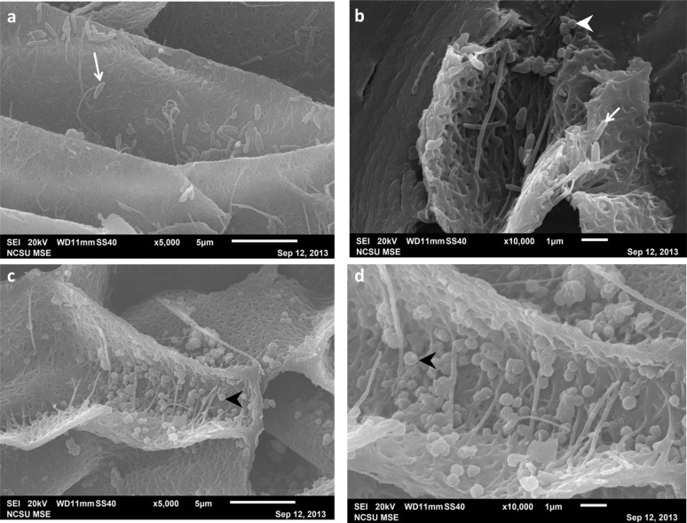
Figure 3. SEM images of melanosomes from extant Gallus gallus feathers.
Black (a,b) and brown (c,d) chicken feathers sectioned longitudinally. (b) Although the feather is visually black, both ovate eumelanosomes (arrow) and round phaeomelanosomes (arrowhead) are present. (c) Phaeomelanosomes (arrowhead) are also observed in the brown feather. (d) Melanosomes exhibit unsmooth, granular surfaces. Using this method, like those observed in TEM, melanosomes appear randomly oriented, rather than in dense mats as reported for fossils (see text). Considerable size variation (e.g. ~.5 μm–~2 μm for the eumelanosomes) is observed between all melanosomes.

