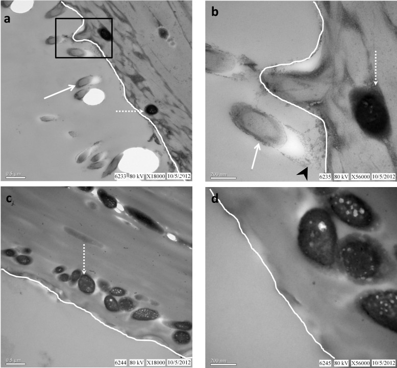
Figure 4. TEM images of Bacillus cereus-treated black chicken feather.
Stained (a,b) and unstained (c,d) TEM images of feather are compared. Feathers were incubated with cultured B. cereus for three days (See SI for details). (a) Superficial B. cereus cells (arrow) extending from the barb surface (white line; feather tissue is to the right). Melanosomes (dashed arrow) are always internal to the feather surface, sparsely distributed and non-overlapping. (b) Higher magnification of boxed area in (a) shows interaction (arrowhead) of bacteria (arrow) with barb surface (white line). White dashed arrow depicts internal, electron-opaque melanosome. Without staining, (c) bacteria are not visible on the external surface, as they are not normally electron-opaque, in contrast with easily visualized, internal, electron-opaque melanosomes (dashed arrow). The melanin pigment is inherently electron dense; no staining is necessary. (d) Enlarged image of unstained section in (c) shows vacuoles associated with internal melanosomes, as has been noted previously4. Keratinous matrix completely surrounds melanosomes, making them difficult to image in SEM without additional treatment.

