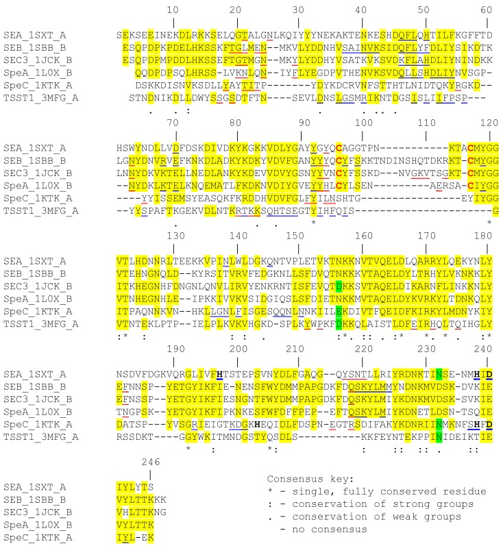
Figure 1.
Sequence alignment of various superantigen sequences. The sequence of each superantigen (SAg) was obtained from the PDB file corresponding to its crystal structure. Multiple sequence alignment [CLUSTAL W (1.81)] was performed using “Biology WorkBench” online tool. Positions with homologous amino acids in three or more SAg sequences are highlighted in yellow or green. Residues in red are involved in forming the characteristic disulfide loop in certain SAg. Residues underlined in red and in blue are involved in binding to Vβ and class II MHC, respectively. Residues in bold are involved in binding zinc.

