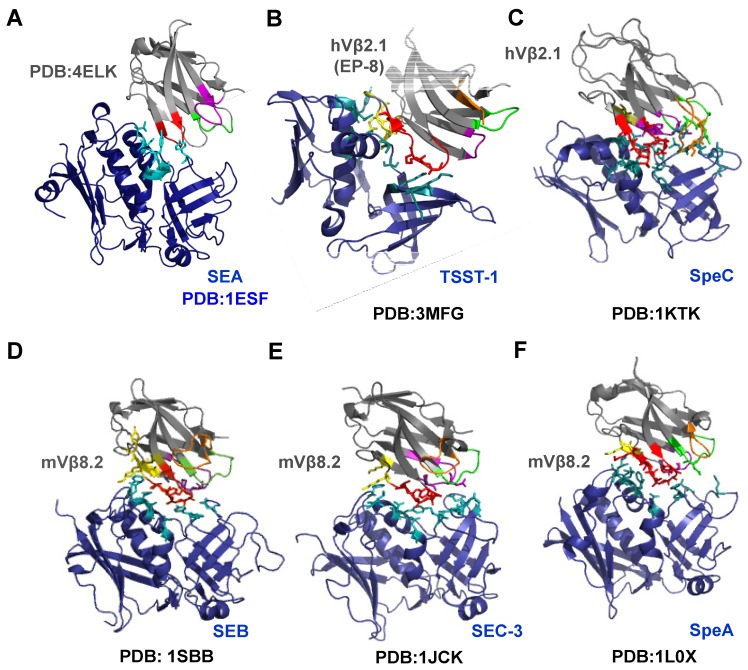
Figure 5.
Co-crystal structures of six superantigens with cognate Vβ domain of the T cell receptor. Except for SEA, the co-crystal structures of SAg (blue) with their cognate Vβ ligand (gray) are available in PDB (Table 1). Residues of SAg interacting with Vβ are indicated in teal. Various regions of Vβ are colored as follows: CDR1 (green); CDR2 (Red); CDR3 (orange); HV4 (purple); FR2 (olive) and FR3 (yellow). Interacting residues of Vβ and SAg are displayed in stick configurations. (A) The SEA crystal structure was manually docked with mouse Vβ16 crystal structure (PDB: 4ELK), that is 66% identical to human Vβ22 protein sequence; (B) Co-crystal structure of TSST-1 with human Vβ2.1 mutant, EP-8; (C) Co-crystal structure of SpeC with human Vβ2.1; (D) Co-crystal structure of SEB with mouse Vβ8.2; (E) Co-crystal structure of SEC3 with mouse Vβ8.2; (F) Co-crystal structure of SpeA with mouse Vβ8.2.

