
Figure 7. Stent Surface Endothelial Cell Proliferation Is Impaired to the Greatest Extent in Rabbits Treated With ZES-Mf.
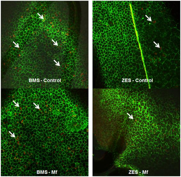
Confocal microscopy was conducted on 14-day whole-mount stented (BMS or ZES) arterial segments in the presence or absence of Mf (100 mg/kg/day) using dual immunofluorescence for platelet endothelial cell adhesion molecule 1/CD31 (green) and BrdU (red). Representative figures are shown at ×100 magnification. White arrows indicate examples of proliferating endothelial cells. Abbreviations as in Figures 1 and 6.