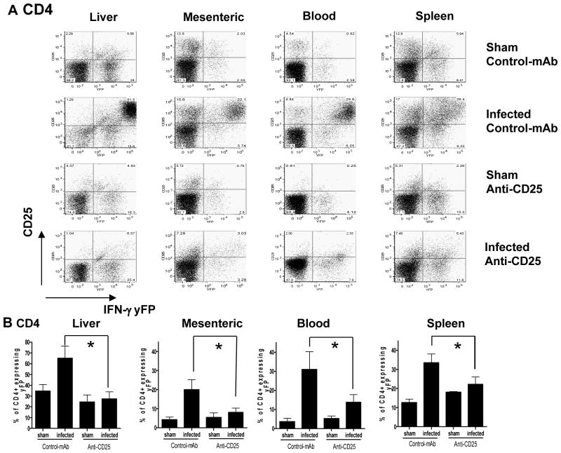
Figure 5. Anti CD25 antibody administration suppresses IFN-γ production by CD4+ T cells.
Groups of 3 bicistronic IFN-gamma-enhanced yellow fluorescent protein (IFN-gamma-YFP) reporter mice (Yeti) male mice were given 1 mg anti-CD25 (PC61) or control mAb (HRPN) i.p 5 days before oral infection with brain homogenate containing10 ME49 cysts or sham uninfected brain homogenate. (A) Representative dot plots showing yFP expression relative to CD25 expression (utilizing 7D4 to detect) on CD4+ T cells from spleen, mesenteric lymph, liver and blood on day 8 post-infection. (B) Relative frequencies of CD4+ T cells expressing yFP in anti-CD25 antibody treated and control antibody treated T. gondii infected and sham infected mice on day 8 post-infection. The results shown are the mean ± std. dev. of the group (n = 3) and are representative of 2 independent experiments. * (p<0.05).

