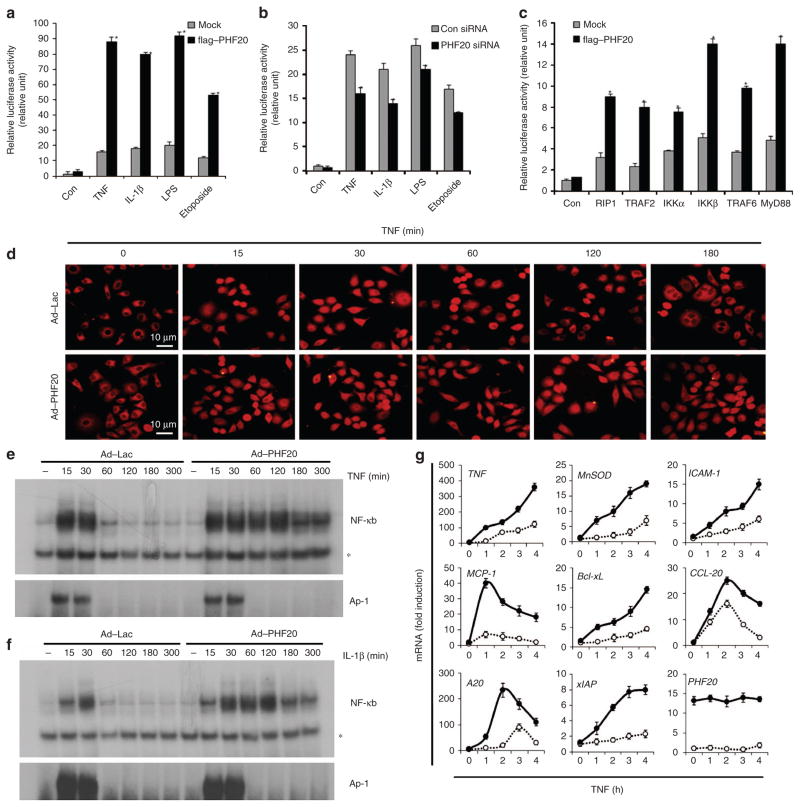
Figure 1. PHF20 is a positive regulator of NF-κB signalling.
(a,b) 293/IL-1R/TLR4 cells were transiently transfected with p2xNF-κB-Luc and pRSV-β-gal with expression plasmids of the flag–PHF20 or PHF20 short interfering RNA (150 pmol). After 24 h of transfection, cells were treated for 6 h with TNF (15 ng ml −1), IL-1β (10 ng ml −1), LPS (1 μg ml −1) and etoposide (20 μM). (c) 293/IL-1R/TLR4 cells were transiently transfected with indicated expression plasmids along with p2xNF-κB-Luc and pRSV-β-gal. Luciferase assays were performed as described in Methods, and the activity of each sample was normalized according to β-galactosidase activity. Each columns shows mean±s.e. of at least three independent experiments. *P<0.05, compared with mock-transfected cells (Student’s t-test) (d–g) HeLa cells were infected with either rAd–Lac or rAd–PHF20 for 24 h, and then treated with TNF or IL-1β for indicated times. (d) Subcellular localization of p65 was analysed by immunofluorescence confocal microscopy with a rabbit anti-p65 antibody (red). Scale bar, 10 μM. (e, f) Electrophoretic mobility shift assay was performed as described in Methods. AP-1-binding activity serves as a positive control. * indicates nonspecific binding to NF-κB probe. (g) mRNA expression levels of NF-κB target genes (TNF,MnSOD,ICAM-1,MCP-1,Bcl-xL,CCL-20,A20 and xIAP) were measured by real-time RT–PCR (○, rAd–Lac; ●, rAd–PHF20). The data represent results from three independent experiments (mean±s.e. of triplicate).

