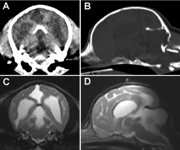
Fig. 1.

CT and MR images of the cranium and brain. A transverse CT image (A) showing bilateral ventriculomegaly and a cystic structure that occupied the region where the cerebellar vermis is normally found. The cystic structure extended into the occipital lobe. On a reformatted parasagittal CT image (B), dorsal displacement of the rostral cerebellar tentorium and a small defect in its central portion were observed. Part of the left condyle of the occipital bone protruded into the caudal fossa. On transverse and midsagittal T2-weighted MR images (C and D), the dilation of the fourth ventricle and the presence of a large cystic structure in the region that normally contains the vermis were confirmed. The cyst extended supratentorially and had displaced the occipital lobes. The MRI signal characteristics of the cystic structure were indicative of fluid accumulation.
