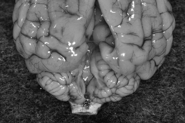
Fig. 2.

A dorsal view of the postmortem brain specimen. Following the removal of the fluid-filled cyst, we found that the cerebellar vermis was absent. The cerebellar hemispheres were normal in size. The occipital lobes exhibited indentation caused by compression by the cyst.
