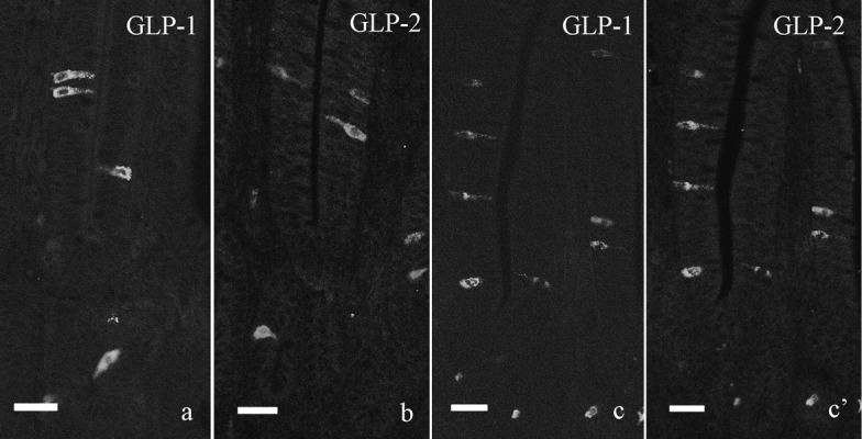
Fig. 1.

Immunofluorescent staining for GLP-1 (a, c) and GLP-2 (b, c’) in the chicken distal ileum. a, b: Single immunofluorescent staining for GLP-1 (a) and GLP-2 (b). Both immunoreactive cells are scattered in villous epithelium and crypt of the distal ileum and show the similar localization to that indicated by double immunofluorescent staining. c, c’: Double immunofluorescent staining for GLP-1 (c) and GLP-2 (c’). Most GLP-1-immunoreactive cells also show immunoreactivity for GLP-2. Bar=20 µm.
