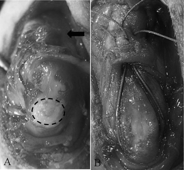
Fig. 4.

Intraoperative views of the left elbow joint. Careful removal of the fibrous sac and serous fluid allows visualization of the avulsed tendon end (arrow) and proximal olecranon (dotted line) (A). Two horizontal mattress sutures using polyester are placed through the triceps tendon and two tunnels drilled in the olecranon to reattach the tendon to the proximal olecranon (B).
