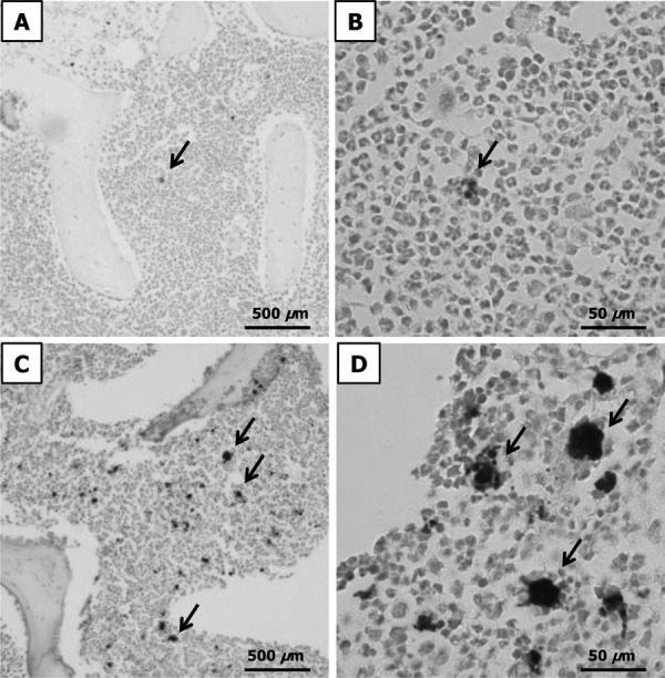
Fig. 5.

Histopathological changes in bone marrow tissue sections, analyzed using Prussian blue staining. The arrows show hemosiderin. A, B: low- and high-power light microscopy images for Day 0. C, D: low- and high-power light microscopy images for Day 42.
